
The Most Unusual Things Scientists Have Found Inside Living Cells
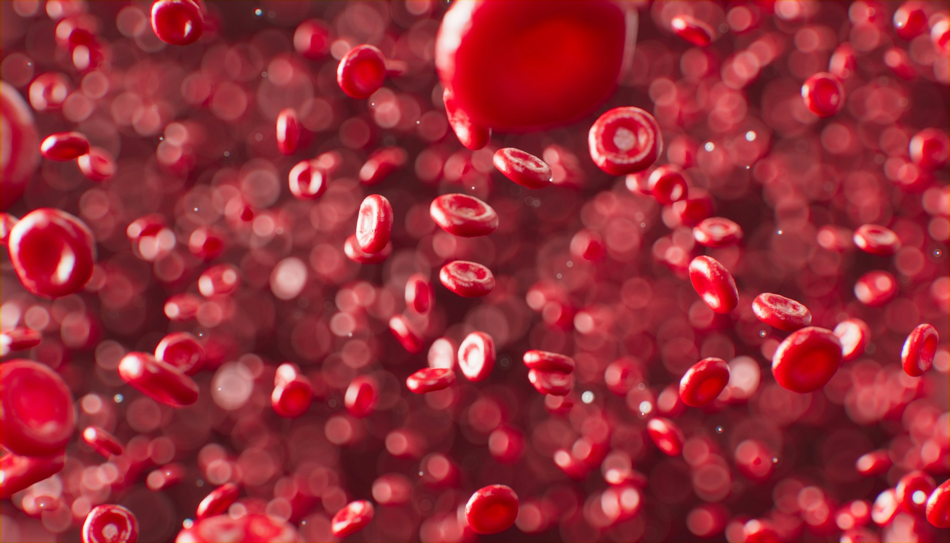
There is a universe hiding inside you, and it does not require a telescope to explore.
It is small enough to fit inside a microscopic cell, yet complicated enough to contain molecular machines, transportation networks, chemical factories, recycling systems, electrical signals, and instructions for building an entire organism. Some of its components are so strange that, if they were discovered floating somewhere in deep space, scientists might mistake them for the remains of an unfamiliar civilization.
We often imagine cells as tiny bags of liquid containing a nucleus and a few scattered biological components. Textbooks sometimes simplify them into neat diagrams, with mitochondria shaped like beans and chromosomes arranged like carefully organized strings. But real cells are far more dynamic, crowded, and surprising than those illustrations suggest.
Scientists have discovered structures that assemble and disassemble according to changing conditions, molecular machines that walk along microscopic tracks, compartments that appear without traditional membranes, and remnants of ancient viral encounters preserved inside cellular machinery.
Some cells even contain structures that challenge our ordinary understanding of what it means for something to be alive.
The more researchers investigate these microscopic worlds, the more they discover that a cell is not simply a building block of life. It is an entire environment, filled with activity that rarely stops.
And some of the most unusual things inside it are hiding in plain sight.
1. Molecular Machines That Walk Through the Cell
Imagine a microscopic worker carrying a package across a crowded factory floor. It follows a designated route, takes carefully measured steps, and delivers its cargo to a particular destination.
Inside living cells, something remarkably similar happens.
Proteins called kinesins and dyneins transport materials through the cell using structures known as microtubules. These microscopic tracks form part of the cell’s internal skeleton, providing both structural support and pathways for transportation.
Kinesin proteins commonly move toward one end of a microtubule, while many dyneins move toward the opposite end. Their movements help transport vesicles, proteins, and other cellular cargo.
What makes this extraordinary is that these machines do not possess legs in the familiar biological sense. They use changes in their molecular structure, powered by chemical energy from ATP, to move along their tracks.
A protein is not consciously deciding where to go. Its movement emerges from chemistry, molecular shape, and interactions with its surroundings.
Nevertheless, the result resembles a sophisticated logistics system.
Different materials must reach different destinations. Some proteins need to travel toward the cell’s outer regions, while other cargo must move toward the interior. In neurons, where extensions can stretch over considerable distances, transporting materials efficiently is especially important.
A cell therefore contains an elaborate transportation network operating at a scale where ordinary mechanical engineering gives way to molecular physics.
The surprising lesson is that movement does not require muscles, wheels, or visible machinery. At sufficiently small scales, a collection of interacting molecules can perform work with remarkable precision.
2. Liquid-Like Droplets That Organize Important Cellular Activities
For a long time, researchers commonly pictured the inside of a cell as a collection of membrane-bound compartments suspended in a fluid.
That picture remains useful, but it does not tell the entire story.
Scientists have discovered that some cellular components gather into temporary, concentrated regions without being enclosed by conventional membranes. These structures are often called biomolecular condensates.
They can form when proteins, RNA, and other molecules interact in ways that cause them to separate from their surroundings, much as oil and water form distinct regions under certain conditions.
Some condensates behave like liquid droplets. Others are more gel-like or have more complicated physical properties.
One familiar example is the nucleolus, a structure inside the nucleus where much of the machinery for producing ribosomal RNA and assembling ribosome components is concentrated.
Other condensates help organize RNA processing, regulate gene expression, or influence how cells respond to stress.
What makes these structures particularly interesting is that they can change over time. Molecules enter and leave them, their composition shifts, and their formation can respond to changing cellular conditions.
There is no need to build a permanent wall around every biological process. Sometimes, the chemistry of the molecules themselves creates a temporary workspace.
This discovery has encouraged scientists to think differently about cellular organization. A cell can create functional neighborhoods through molecular interactions alone.
It is a little like a busy room in which people naturally gather into separate groups to discuss different subjects. Nobody has constructed walls between the groups, yet the arrangement still creates a form of organization.
The comparison is imperfect, but it captures something important: order can emerge without every component being physically separated from everything else.
3. Tiny Biological Compartments That Break the Rules
Not every structure inside a cell fits comfortably into the categories we expect.
Consider the bacterial microcompartment, a specialized structure found in certain bacteria. It resembles a microscopic container assembled from proteins rather than the lipid membranes that surround many familiar cellular compartments.
These structures enclose particular enzymes and help organize specific biochemical reactions.
Some bacterial microcompartments participate in the processing of carbon compounds. Their protein shells help manage the local chemical environment, including the movement of particular molecules into and out of the compartment.
Another example is the carboxysome, a bacterial structure involved in carbon fixation. It concentrates enzymes that help convert carbon dioxide into forms the organism can use to build biological material.
By gathering the necessary components into a confined space, a carboxysome can improve the efficiency of important reactions.
The remarkable part is that these compartments are constructed from protein building blocks that assemble into organized shells.
They are not miniature organs in the conventional animal sense, but they perform specialized functions within the cell.
This is an important reminder that biology does not rely on a single architectural blueprint.
Evolution has produced multiple ways to organize chemical activity. Some systems use membranes, some use protein shells, and others depend on temporary molecular assemblies.
The microscopic world is full of different solutions to the same engineering problem: how do you make a complicated chemical process work reliably in a crowded environment?
4. Cells Contain Their Own Recycling Facilities
Every living cell must manage waste.
Proteins become damaged. Molecular components stop functioning correctly. Structures are no longer needed. Nutrients must be recovered from materials that have already served their purpose.
Cells cannot simply discard everything and start over. Instead, they possess sophisticated systems for dismantling and reusing biological components.
One of the most fascinating is the proteasome, a large molecular complex that breaks down proteins marked for destruction.
The proteasome resembles a barrel-shaped machine with a controlled entrance. Proteins selected for degradation are unfolded and fed into its central chamber, where they are cut into smaller peptide fragments.
Those fragments can then be processed further, and their constituent amino acids may be reused to build new proteins.
Another major recycling system is the lysosome, a membrane-bound compartment containing enzymes that break down various biological materials. Lysosomes help digest damaged cellular components, ingested substances, and other material that needs to be processed.
A related process called autophagy allows cells to deliver portions of their own contents to lysosomes for degradation and recycling.
During certain forms of autophagy, a membrane grows around material destined for breakdown, eventually forming a compartment that fuses with a lysosome.
The contents are then dismantled, allowing useful components to be recovered.
Think of it as a factory that can disassemble its own equipment, recover the raw materials, and use them to manufacture replacements.
This is not merely a clever convenience. Cellular maintenance is essential to survival. The ability to identify damaged components, remove them, and recover valuable resources helps organisms respond to changing conditions.
Life is not just about building things.
It is also about knowing what to take apart.
5. Mitochondria Have Their Own DNA
Mitochondria are often described as the powerhouses of the cell because they play a central role in producing ATP, a molecule that supplies usable energy for many biological processes.
But their history is stranger than their familiar nickname suggests.
Mitochondria contain their own genetic material, separate from the DNA stored in the cell nucleus.
In humans, mitochondrial DNA is a small circular molecule containing genes essential to mitochondrial function. It represents only a tiny fraction of the genetic information found in a human cell, but it has important biological roles.
Why would an organelle responsible for energy production have its own DNA?
The leading explanation comes from the theory of endosymbiosis. According to this account, mitochondria evolved from bacteria that entered into a long-term relationship with an ancestral cell billions of years ago.
Rather than remaining independent organisms, these bacteria became integrated into the biology of their host. Over evolutionary time, many of their genes were lost or transferred to the host’s nuclear genome.
The descendants of this ancient partnership became the mitochondria found in modern eukaryotic cells.
Mitochondria still retain features that reflect this history, including their own genetic material and bacterial-like ribosomes.
They are not independent organisms living freely inside us. They depend extensively on the cells that contain them. Nevertheless, their evolutionary origins are written into their structure.
Every time a human cell produces energy using mitochondria, it relies on the descendants of a partnership that began in a world vastly different from our own.
It is one of biology’s most remarkable examples of how cooperation can transform the organization of life.
6. Cells Contain Structures That Change Shape in Seconds
A cell may appear stationary under a microscope, but its interior is anything but static.
Many cellular structures continuously assemble, change shape, and disappear as conditions change.
One striking example is the actin cytoskeleton, a network of protein filaments that helps cells maintain their shape, move, and organize their internal components.
Actin filaments can grow when individual protein subunits are added and shrink when subunits are removed. By regulating these processes, cells can reorganize their internal architecture.
Some cells use this ability to crawl across surfaces. Others use it to change shape, divide, or form specialized structures.
During cell division, another dynamic structure becomes visible: the mitotic spindle.
Made primarily from microtubules and associated proteins, the spindle helps separate duplicated chromosomes so that each daughter cell receives the appropriate genetic material.
The spindle is not a rigid mechanical device that simply remains in place while chromosomes move through it. Its microtubules continually grow and shrink, attach to chromosomes, and reorganize as division proceeds.
The cell coordinates these dynamic activities through molecular interactions and regulatory systems.
What looks like a stable structure in a textbook illustration may actually be a temporary arrangement of components constantly being replaced.
This reveals a subtle principle of living systems: stability does not always require stillness.
A river maintains its overall shape while its water flows. A flame persists while fuel and gases continually move through it. In a different and more complicated way, certain cellular structures preserve their functions while their molecular components remain in motion.
The cell is full of structures whose identities depend less on having permanent parts than on maintaining an organized pattern of activity.
7. Some Cells Carry Ancient Viral Remnants
Not everything unusual inside a cell is a structure built for an obvious purpose. Some of the most surprising molecular features are remnants of encounters that occurred during evolution.
Viruses have interacted with living organisms for an immense span of evolutionary history. Certain viral sequences have become incorporated into the genomes of their hosts.
When such sequences enter the reproductive lineage of an organism and are inherited by its descendants, they can persist across many generations.
These inherited sequences are called endogenous viral elements. In humans and other mammals, a substantial collection of genomic sequences originated from ancient retroviral infections.
Most of these sequences no longer function as complete viruses. Many have accumulated mutations that prevent them from producing infectious viral particles.
However, some have acquired or retained biological functions.
A particularly striking example involves genes derived from ancient retroviruses that contribute to the formation of the placenta in many mammals.
In humans, the proteins syncytin-1 and syncytin-2 participate in processes associated with the fusion of cells involved in placental development. Their evolutionary origins are connected to ancient retroviral envelope proteins.
In other words, molecular machinery derived from ancient viral genes became part of an essential biological process.
This does not mean that a human cell is secretly filled with active viruses. The important distinction is that genetic material left behind by ancient infections can sometimes be repurposed over evolutionary time.
Evolution does not always work by inventing entirely new components. Sometimes it modifies existing ones, including material that originated in circumstances very different from those in which it is used today.
The result can be something unexpected: a biological feature whose history includes a virus, but whose present-day function contributes to the development of an organism.
8. Cells Can Build Electrical Gradients Across Their Membranes
Electricity is often associated with wires, batteries, and electronic devices. Yet living cells also depend on electrical differences, although their mechanisms are based on ions moving across membranes rather than electrons flowing through a metal wire.
The inside and outside of a cell typically differ in their concentrations of charged particles, including sodium, potassium, calcium, and chloride ions.
Because the cell membrane regulates the movement of these ions, an electrical potential develops across it.
This is called the membrane potential.
In many animal cells, the membrane potential is maintained partly through ion channels and transport proteins, including the sodium-potassium pump.
The pump uses energy from ATP to move sodium and potassium ions in opposite directions across the membrane, helping maintain the concentration differences that contribute to the electrical potential.
In neurons, changes in membrane potential allow electrical signals to travel along the cell. In muscle cells, electrical activity helps trigger contraction. Other cells use electrical and ionic changes to regulate secretion, transport, and biochemical activity.
The fascinating part is that a cell can store and manipulate electrical potential without containing any metal wires or conventional electronic components.
Its boundaries become part of an electrical system.
The cell membrane is not simply a wall that keeps the contents inside. It is an active interface that regulates the movement of matter and helps establish the physical conditions necessary for life.
In some organisms, groups of cells coordinate electrical activity in ways that influence development, movement, and responses to the environment.
Biology and electricity are therefore not separate subjects. They meet at the boundary of almost every living cell.
9. Some Cells Contain Crystals and Mineral Structures
Living cells are often associated with soft tissues, flexible membranes, and watery interiors. But biological systems can also create remarkably ordered mineral structures.
One example is the formation of magnetosomes in certain bacteria.
These specialized structures contain magnetic mineral crystals, commonly magnetite or greigite, enclosed within membranes. The crystals are arranged in ways that allow the bacteria to respond to magnetic fields.
For many magnetotactic bacteria, this helps orient their movement along Earth’s magnetic field, guiding them toward environments with suitable oxygen concentrations and other conditions.
The result is an organism whose internal structures include tiny biological magnets.
Another example appears in organisms that produce mineralized structures for protection, support, or specialized functions. Some single-celled organisms create intricate shells or skeletons from minerals, while certain microorganisms accumulate mineral deposits within or around their cells.
In humans, mineral formation is essential to the development of bones and teeth, although these are structures formed through the coordinated activity of many cells rather than isolated intracellular crystals alone.
The surprising principle is that living systems can control the formation of solid materials with considerable precision.
Proteins and other biological molecules influence where minerals form, how crystals grow, and what shapes they adopt.
This process, known broadly as biomineralization, connects biology with materials science.
Scientists study it not only to understand organisms but also to investigate possible approaches to producing materials under relatively mild conditions.
Nature has spent billions of years exploring ways to build structures from available ingredients. Some of those solutions are so unusual that they continue to inspire modern research.
10. The Cell Nucleus Is Not an Organized Library of Perfectly Sorted Books
The nucleus contains the genetic instructions needed to maintain and regulate a eukaryotic cell. It is tempting to imagine that its DNA is arranged like a library, with every gene stored neatly in its own labeled section.
The reality is more dynamic.
DNA is wrapped around proteins called histones, forming a material known as chromatin. Chromatin folds and organizes itself into structures that influence how genes can be accessed.
Some regions are relatively open and associated with active gene expression. Others are more compact and generally less accessible.
The genome also occupies a three-dimensional arrangement inside the nucleus. Different regions of DNA can come into contact, bringing regulatory elements close to genes that may be located far away along the DNA sequence.
Scientists use techniques such as chromosome conformation capture to investigate these spatial relationships.
They have discovered that chromosomes form organized domains and larger-scale structures that help shape the regulation of genetic activity.
However, these arrangements are not permanent. Chromatin organization changes with cellular activity, development, and other conditions.
A gene’s behavior can depend not only on its DNA sequence but also on its regulatory environment, including the proteins associated with the DNA and the way the surrounding chromatin is organized.
The nucleus is therefore not simply a storage container.
It is a dynamic environment in which genetic information is packaged, accessed, regulated, and maintained.
The instructions remain encoded in the DNA, but the way a cell uses those instructions depends on a much larger system of molecular relationships.
11. Some Cells Contain Entire Communities of Microorganisms
A particularly surprising discovery emerges when we stop thinking about a cell as an isolated biological unit.
Certain organisms maintain close relationships with microorganisms that live inside their cells. These relationships can be so integrated that the host depends on its microscopic partners for essential biological functions.
For example, some insects contain specialized cells that house bacterial symbionts.
Aphids commonly rely on bacteria called Buchnera aphidicola, which live inside specialized host cells known as bacteriocytes. These bacteria provide essential amino acids that are limited in the aphids’ plant-based diet.
The relationship is not a casual encounter between two independent organisms. It is a highly integrated partnership shaped by a long history of coevolution.
The host provides a protected environment and nutrients, while the bacteria contribute compounds that help the host survive.
Other examples occur in marine organisms, including certain animals that maintain symbiotic bacteria involved in nutrition or chemical processes.
Some of these partnerships are so specialized that the host and its microorganisms depend heavily on one another.
Such relationships blur the intuitive boundary between an individual organism and its biological partners.
A living system can sometimes be understood only by examining the interactions between its different components, even when those components belong to distinct evolutionary lineages.
The cell is not always a solitary factory manufacturing everything it needs. In some cases, it is also a habitat.
12. There Are Tiny Cellular Structures That Help Protect Against Viruses
Bacteria and archaea live in environments where viruses can be abundant. To survive, many have evolved molecular systems that recognize and respond to viral genetic material.
One of the best-known examples is CRISPR-Cas immunity.
Some microorganisms preserve fragments of genetic material from previous viral encounters within specialized regions of their genomes called CRISPR arrays.
These fragments can be transcribed into RNA molecules that help guide associated Cas proteins toward matching genetic sequences.
When a related virus attacks again, certain CRISPR-Cas systems can recognize and cut the invading genetic material.
The precise mechanisms differ among CRISPR systems, and not every system works in exactly the same way. Nevertheless, the central concept is extraordinary: a microorganism can preserve molecular records of past encounters and use them to help recognize future threats.
The process does not involve conscious memory. It is a biochemical system in which stored genetic sequences and specialized proteins work together.
Researchers discovered that these natural defense mechanisms could be adapted into powerful tools for editing genetic material, transforming fields of biological research.
The original function, however, emerged within microorganisms that needed ways to survive in a world full of viruses.
A tiny cell, with no brain and no nervous system, can possess a molecular defense system that reflects elements of its biological history.
The microscopic world is full of such surprises: complex functions emerging from chemical interactions and evolutionary processes rather than from centralized control.
13. Some Cellular Structures Are Built From RNA, Not Just Proteins
RNA is often introduced as a messenger that carries genetic instructions from DNA to the machinery responsible for making proteins.
That description is useful, but incomplete.
RNA can also perform structural and catalytic roles.
One of the most important examples is the ribosome, the molecular machine that builds proteins by linking amino acids together according to instructions carried by messenger RNA.
Ribosomes contain both proteins and ribosomal RNA, or rRNA. Although proteins are important to ribosome structure and function, the catalytic center responsible for forming peptide bonds is primarily made from RNA.
This makes the ribosome an example of a ribozyme: a molecular structure in which RNA performs a catalytic function.
Other RNAs help regulate genes, process RNA molecules, and guide proteins to particular targets.
Some RNA molecules form elaborate three-dimensional shapes that determine how they interact with other molecules.
These discoveries challenged the simple assumption that proteins are the only biological molecules capable of carrying out sophisticated chemical work.
RNA can store information, interact selectively with other molecules, and catalyze reactions.
This versatility has also contributed to scientific investigations into the origins of life, including hypotheses that early biological systems may have relied more heavily on RNA before the evolution of modern DNA-and-protein-based systems.
That possibility remains an area of scientific investigation, but the capabilities of modern cellular RNA are already remarkable.
A molecule commonly associated with carrying messages turns out to be capable of helping build the machinery that reads those messages.
The messenger is part of the machine.
The Microscopic World Is Stranger Than It Looks
The most unusual discoveries inside living cells share a common feature: they reveal that life does not depend on a single kind of organization.
Cells use molecular machines that walk along microscopic tracks. They create temporary compartments without conventional membranes. They recycle damaged components, maintain electrical gradients, construct mineral structures, and preserve genetic remnants of ancient biological encounters.
Their interiors contain systems that are simultaneously physical, chemical, and informational.
A protein’s shape determines what it can bind. An electrical gradient influences how ions move. The arrangement of DNA affects which genes can be accessed. The organization of molecules can create compartments that appear and disappear as conditions change.
None of these processes requires a tiny conscious operator sitting inside the cell.
Instead, biological organization emerges from the interactions of countless molecules, governed by physical laws and shaped by evolutionary history.
That does not make the cell less extraordinary. It makes the explanation more interesting.
A microscopic structure can perform a function that appears mechanical. A molecule can carry information across generations. A collection of proteins can form a temporary workspace. A genetic remnant from an ancient virus can eventually contribute to an essential biological process.
The cell is not a miniature version of a human-built machine. It is a different kind of system, one that continually maintains itself, responds to changing conditions, and uses its own components to repair and reorganize its internal environment.
And we have only begun to understand the full complexity of that world.
Every improvement in microscopy, molecular analysis, and computational biology creates opportunities to investigate structures that were previously invisible or impossible to characterize.
Scientists can now observe molecular interactions, reconstruct cellular organization, and investigate processes that once existed only as hypotheses.
Some discoveries will refine what we already know. Others will reveal entirely new ways in which living systems organize themselves.
The next extraordinary discovery might be inside a bacterium living in the soil, a neuron carrying a signal through the brain, or a single cell dividing beneath a microscope.
We do not need to travel to another planet to find a world full of unfamiliar structures and remarkable processes.
Sometimes, the most unusual universe is already alive.
It is simply too small to see without the right tools.